How to Draw a Cell Diagram
I'g a instructor with more than a decade of experience in science pedagogy.
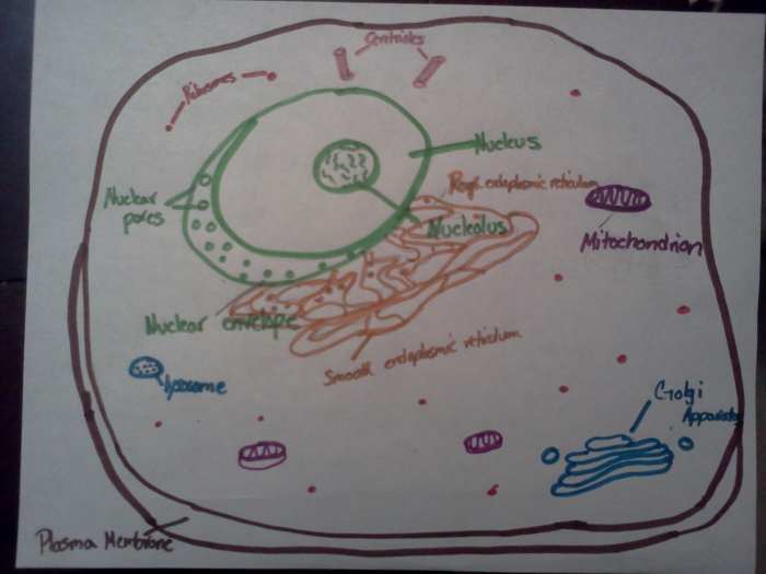
My simple drawing of an brute prison cell.
Patrice M
Instruction most cells was one of my favorite units. Especially in lower grades when students demand to just know the basics, there are many fun activities to practise.
Drawing cells is typically not a skill assessed on tests or required by standards, but information technology can certainly assistance students develop a lasting knowledge of the cell. I would never practice this in isolation but rather aslope learning about the construction and function of parts of an animal jail cell.
Hither is a tutorial with pictures demonstrating how to draw an creature cell. I am non an artist—so if I can exercise it, anyone can.
Plasma Membrane
The plasma membrane is a flexible membrane that covers all cells. It allows sure materials in and out of the cell which makes it semi-permeable. In animal cells, this is the only covering between the inside and outside of the jail cell so it gives information technology a round or fluid shape.
Cell Construction
The starting time thing you want to conspicuously illustrate in your drawing is the exterior construction of the animal cell. Although the jail cell is 3 dimensional and your cartoon will be two dimensional, there are techniques to show dimension in your drawing. Simply add an additional line around two sides of the exterior of the brute prison cell. And then, characterization this the plasma membrane.
Summary of Cell Organelle Functions
| Organelles | Function |
|---|---|
| Centriole | Occur in pairs and important for cell sectionalisation |
| Endoplasmic reticulum | A highly folded membrane that is that site for poly peptide and lipid synthesis |
| Golgi Apparatus | A flattened stack of membranes that modifies proteins and packages them in the cell |
| Lysosome | A vesicle that contains digestive materials to break down cellular wastes |
| Mitochondrion | A membrane- leap organelle that makes energy available to the remainder of the cell |
| Nucleus | Control centre of the prison cell that contains directiosn for the production of proteins and cell partition |
| Plasma membrane | A flexible boundary that controls the movement of substances int oand out of the cell |
| Ribosome | Synthesize proteins |
| Vacuole | A membrane bound vesicle that stores nutrient and water |
The Nucleus
The nucleus is the control center of the cell and accordingly a large structure in the cell. It manages the activities of the residual of the cell. Information technology contains Deoxyribonucleic acid which contains the information needed to make proteins necessary for growth and reproduction. The nucleus has its own membrane that has pores allowing things to leave the nucleus.
Inside the nucleus is another structure called the nucleolus where ribosomes are produced. As yous draw the nucleus, create aforementioned illusion of a membrane equally the one created for the plasma membrane surrounding the prison cell. Also include lines for Dna and a round structure for the nucleolus.
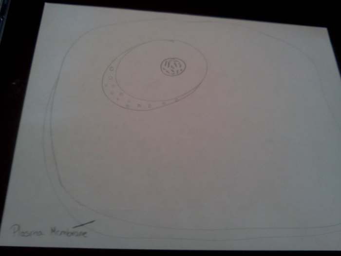
The cell membrane showing that this is a cantankerous department drawing. Too, notice the size of the nucleus in rlation to the rest of the jail cell.
Patrice 1000
Endoplasmic Reticulum
Next, we accept the endoplasmic reticulum (en duh PLAZ mihk - rih TIHK yum lum) or ER for short. Agreement the construction and location of the endoplasmic reticulum will assist you empathize its function. The ER is a arrangement of highly folded membrane sacs and interconnected channels where protein and lipid synthesis occur. Information technology is fastened to the nucleus because ribonucleic acrid (RNA) transcribed in the nucleus travels out of nuclear pores and onto the ER to translate proteins. The many folds of the ER provide more surface area for ribosomes to produce proteins. The part of the ER that contains ribosomes is called the Rough ER.
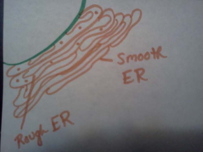
The Smooth and Rough ER are attached to the nucleus.
Patrice M
Smooth ER
The role of the endoplasmic reticulum that does not contain ribosomes is called the Polish ER. Information technology extends from the rough ER and continues the folds of the rough ER. The Smooth ER is where lipids and complex carbohydrates important for cellular role are fabricated. Phospholipids which make up the jail cell membrane are synthesized in the Smooth ER. Also, Smooth ER is found in the liver where it detoxifies harmful substances.
Ribosomes
Ribosomes are small structures surrounded by a membrane that produce proteins. They can be plant floating freely around the cell or attached to the Rough ER. They are composed of poly peptide and RNA and can be drawn every bit pocket-sized circles in your diagram of an animal cell.
Golgi Apparatus
The Golgi Apparatus is a flattened stack of membranes that modifies, sorts and packages proteins into sacs called vesicles. Later proteins are fabricated in the ribosomes on the Crude ER, some are shipped to the Golgi Apparatus for further processing. Similar vesicles compression off the Golgi carrying proteins to the plasma membrane where the vesicles fuse to release proteins into the environment surrounding the cell. My students use to confuse the ER with the Golgi when I showed them the structures independently. Nonetheless, when I started teaching virtually the cell as a whole and allowing students to draw the entire cell, they could run across the Golgi was freestanding and surrounded by vesicles.
Read More From Owlcation
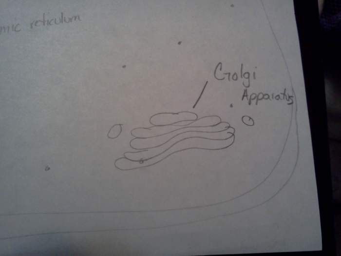
The Golgi is a stack of flattened sacs unremarkably surrounded past vesicles.
Patrice M
Sometimes Vacuoles
Vacuoles are vesicles surrounded by membranes that store food and waste products. Beast cells don't usually comprise vacuoles simply when they do they are small, round structures throughout the cell.
Lysosomes
I always connect Lysol to lysosomes to help my students remember the office. Lysosomes are small vesicles that contain substances for breaking down wastes. Lysosomes tin can digest bacteria and viruses that accept entered the jail cell. Describe your lysosomes like vesicles except include small dots inside them to represent the enzymes that break things down.
Centrioles
Centrioles are structures made of microtubules (like skeleton) that function in cell partitioning. They are commonly near the nucleus because they assist split up the genetic material when the jail cell splits and reproduces. Centrioles are unique to animal cells and look like a bunch of sticks tied together.
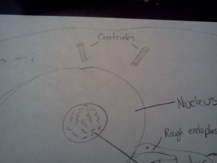
Describe your centrioles in pairs and nigh the nucleus.
Patrice M
Mitochondria
Mitochondria are the free energy generators of a cell. They convert sugar into energy the cell can apply in the grade of ATP. The mitochondria have an outer membrane and a highly folded inner membrane. Just like the ER had folds to increase the surface surface area available this is similar in the mitochondria. The large surface surface area is used for the breaking of bonds in sugars which releases energy for the cell to use. Diagram your mitochondria like beans with a cross department showing the folds of the inner membrane.
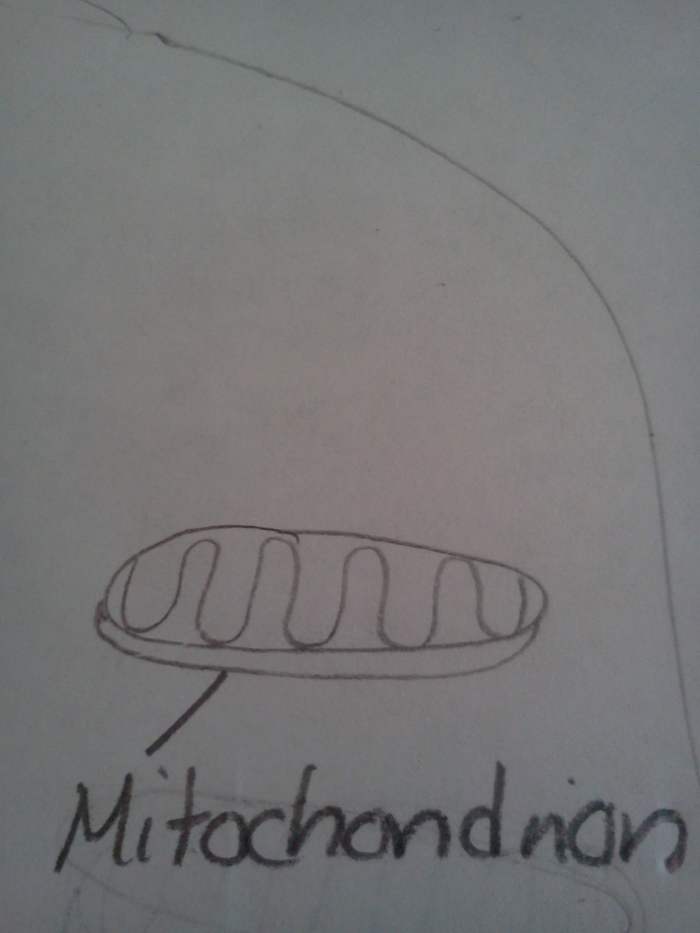
Draw a squiggly line to show the folded inner membrane of the mitochondrian.
Patrice Grand
Those are the chief parts of a cell in an beast that you will have to draw. depending on your grade level yous may add together or remove some structures. Pay attending to showing membranes and relative sizes of the different parts. Of course, learning or reviewing the function while cartoon the structure helps 1 to better sympathise the prison cell. Feel complimentary to add colors to bring out the beauty of your cartoon and your own creativity. Happy Drawing!
vandna on September 17, 2015:
I remember drawing this in my highschool practical books . thanks for sharing . its really cool .
Paul Perry from Los Angeles on April 29, 2013:
wow this is super cool thank you for making the hub!
sociopath from NEW YORK on January 21, 2013:
That was a bit difficult to draw. Thanks for sharing.
Nick DeArcangelis from Illinois on January 21, 2013:
I had to draw cells for Biological science last yr! This probably would have helped immensely equally I was drawing using an already completed picture from the book. Not to mention my drawing skills are... sub-par. Voted up!
frozenink on January 21, 2013:
I recollect coloring it as well! Nice seeing this. Thanks for sharing!
Wakerra on Jan 21, 2013:
aside from the shape, have yous ever played "CellCraft"? Information technology puts the scientific learning experience with the fun cartoony-video game adventure elements
Sheila Craan from Florida on Jan 21, 2013:
Interesting Patrice! I remember seeing less particular in grade schoolhouse and certain learned a lot here! Congratulations!
Wakerra on Jan 21, 2013:
I idea establish cells were square, animate being cells are round
Giani Noyez from Belgium on Jan 21, 2013:
I think having to report these cell structures. It was fairly easy, and when reading this I've had the feeling I still knew everything I've learned.
Nice drawings, with your caption everyone should know what's going on.
JP Carlos from Quezon City, Phlippines on January 21, 2013:
I remember drawing cells in elementary. Nosotros even fabricated a model out of foam, balls and other materials found at domicile. When teachers use creative strategies students will larn the topic faster.
This will make a corking resource for all students out at that place. Swell work and congratulations.
Source: https://owlcation.com/stem/How-to-Draw-a-Cell-from-an-Animal
0 Response to "How to Draw a Cell Diagram"
Post a Comment